BAC Monthly Slide Club Report October 2026
BAC Lunchtime Slide Club explores two challenging cytology cases
The British Association for Cytopathology (BAC) hosted its monthly Lunchtime Slide Club on 28 October 2026, featuring two challenging cases presented by Dr Amy Ly, MD, from Massachusetts General Hospital and Harvard Medical School.
Case 1: Burkitt lymphoma presenting in ascitic fluid
The first case involved a 40-year-old woman with a 30 cm complex pelvic mass, initially suspected to be an ovarian primary tumour. Approximately one litre of ascitic fluid was collected during surgery. The right ovary was replaced by a partially necrotic mass, with associated small-volume aortocaval lymphadenopathy.
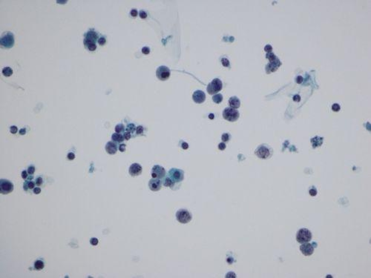
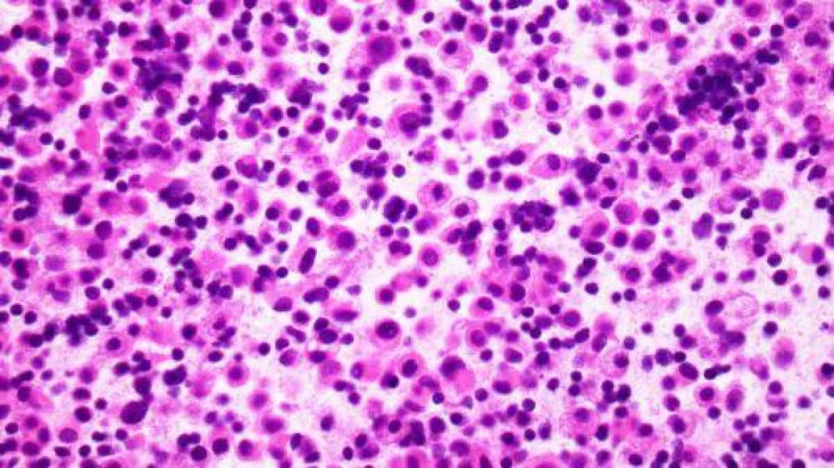
Cytological examination of the ascitic fluid revealed a highly cellular specimen containing relatively discohesive cells with scant cytoplasm, high nuclear-to-cytoplasmic ratios, irregular nuclear contours and prominent apoptotic activity. The degree of degeneration complicated interpretation, with the differential diagnosis including high-grade carcinoma, melanoma, lymphoma and other high-grade neoplasms.
Examination of the cell block supported the presence of a high-grade neoplasm. The ovarian tumour showed positivity for CD20 and BCL6, with a very high Ki-67 proliferation index. Flow cytometry identified a CD10-positive population with lambda light-chain restriction, while cytogenetic findings demonstrated MYC and IGH rearrangements.
Despite some morphological atypia and variation in cell size, the combined morphological, immunophenotypic and genetic findings supported a diagnosis of Burkitt lymphoma.
The case highlighted the importance of recognising the effects of degeneration on cytological interpretation and integrating morphology with ancillary investigations when assessing high-grade lymphoid neoplasms.
Case 2: Lobular carcinoma of the breast identified in pleural fluid
The second case concerned a 68-year-old woman with a significant smoking history who presented with breathlessness and a large unilateral pleural effusion. She also reported pruritus and lower back pain.
The pleural fluid contained a population of predominantly discohesive round cells with moderate amounts of cytoplasm, atypical binucleate forms, prominent nucleoli and occasional cohesive clusters of atypical epithelioid cells. The differential diagnosis included adenocarcinoma, melanoma, lymphoma and mesothelioma.
Immunocytochemical investigation was essential to resolving the case. Claudin-4 staining showed predominantly cytoplasmic granular staining rather than the convincing linear membranous pattern expected for a positive result. Calretinin was negative, and further investigation, including positive GATA3 and oestrogen receptor staining, supported the diagnosis of metastatic lobular carcinoma of the breast.
The patient’s breast was biopsied and showed invasive lobular carcinoma with a pleomorphic component, accounting for some of the more atypical cytological features. The disease was stage IV at presentation, with involvement of multiple sites, including lymph nodes and the pleura. Her lower back pain was associated with pathological spinal fractures.
This case demonstrated the diagnostic challenges of identifying metastatic lobular breast carcinoma in serous fluids, particularly when the primary tumour may be difficult to detect on conventional imaging. It also reinforced the importance of interpreting immunocytochemical staining patterns carefully and using an appropriate panel of markers to distinguish between epithelial and mesothelial lesions.
Key learning points
Both cases demonstrated the value of combining careful morphological assessment with immunocytochemistry, flow cytometry and genetic investigations where appropriate.
The discussion also highlighted the diagnostic pitfalls associated with degenerative changes, unusual tumour morphology and the interpretation of immunocytochemical staining patterns in challenging serous fluid specimens.