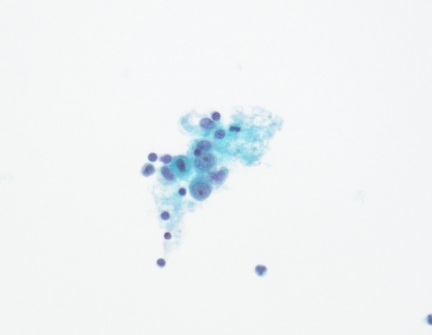
BAC Monthly Slide Club – Challenging Serous Fluid Cytology Cases
Dr Kartik Viswanathan (Emory University, Atlanta) was the invited speaker for the British Association for Cytopathology (BAC) Monthly Slide Club. He presented two challenging pleural fluid cytology cases that highlighted the value of integrating morphology, immunocytochemistry and clinical information when investigating subtle malignant effusions.
Case 1 – Metastatic Renal Cell Carcinoma Presenting as a Pleural Effusion
A 62-year-old man presented with a large right pleural effusion. Initial examination of both the ThinPrep slide and cell block suggested a benign reactive process, with lymphocytes and mesothelial-appearing cells but no obvious malignant population. Standard epithelial markers (BerEP4 and MOC31) were negative, while mesothelial markers were inconclusive.
Further review of the patient's clinical history revealed a previously resected renal tumour together with new liver lesions. This prompted additional immunocytochemistry using PAX8 and PAX2, both of which highlighted a subtle malignant population. Correlation with the patient's history confirmed metastatic clear cell renal cell carcinoma involving the pleural fluid.
Learning points
- Always review the full clinical history, particularly when cytomorphology and initial immunostains do not explain the findings.
- Renal cell carcinoma may be missed by conventional epithelial markers, making PAX8 and PAX2 valuable when there is clinical suspicion.
- Interpret PAX8 carefully, recognising its limitations and the importance of antibody clone selection.
Case 2 – An Unusual Endothelial Neoplasm in Pleural Fluid
A 69-year-old woman presented with a recurrent unilateral pleural effusion that had twice been reported elsewhere as negative for malignancy. Although the cells were subtle and initially resembled reactive mesothelial cells, their high nuclear-to-cytoplasmic ratio and unusual appearance prompted further investigation.
Routine epithelial markers were negative, leading to expansion of the immunocytochemical panel. Strong expression of ERG, FLI1 and CD34 suggested endothelial differentiation. Following discussion with the clinical team, a biopsy was performed and confirmed a malignant neoplasm with endothelial features. Although the precise tumour type could not be fully characterised before the patient's death, the case demonstrated how cytology findings directly prompted definitive tissue investigation.
Learning points
- Trust subtle morphological clues when a cell population does not fit the expected appearance.
- Negative epithelial markers should prompt consideration of alternative lineages rather than prematurely concluding a benign diagnosis.
- Close communication between cytopathologists and clinicians can directly influence patient management and further diagnostic investigation.
Key Messages from the Presentation
- Clinical context is essential when interpreting serous fluid cytology.
- Ancillary immunocytochemistry should be used thoughtfully to answer specific diagnostic questions.
- Rare metastatic tumours and vascular neoplasms can closely mimic reactive mesothelial proliferations.
- Maintaining a healthy level of diagnostic suspicion and investigating unexpected findings can prevent missed malignancies.