BAC Lunchtime Slide Club: Cervical Cytology in Focus 30th July 2025
Case 1 – Donna Rainey (NHS Greater Glasgow & Clyde)
Date: 30 July 2025
Host: Sue Mehew
Organised by: British Association for Cytopathology (BAC)
The fifth BAC Lunchtime Slide Club brought members together for an interactive session on cervical cytology, the first time this topic has featured in the series.
Donna presented a complex case involving a 51-year-old patient on HRT.
The presence of only scanty abnormal cells, including an isolated keratinised "tadpole" form and groups with high nuclear-cytoplasmic ratios, may have been why the case was initially missed by primary screening. Thankfully, this was identified on rapid screening as abnormal and despite a first Checker considering this to be negative, whilst a 2nd Checker believed it showed possible diathesis and isolated high-grade severe dyskaryosis with possible invasion.
The case was finally reported as showing severe dyskaryosis.
Subsequent histology showed CIN3 with a foci of squamous carcinoma (FIGO 1A1).
The discussion highlighted the value of rapid screening and quality control processes.
National Invasive Cancer Audit
- Case reviewed as part of the NICA
- 3 slides to review
- 2013 and 2016 slides were originally reported negative and agreed on NICA review.

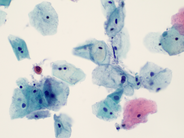
Case 1 from Donna Rainey
2019 Case
Primary Screener – Severe
1st & 2nd Checker – BNC in squamous cells
Medic- BNC in squamous cells
Therefore: False negative on review
Possible reasons:
Scanty cells
Some poorly preserved
There was a discussion about how this should be graded/reported
Case 2019
Case 2 – Sue Mehew (BAC Chair)
Sue shared a case involving a 33-year-old with a history of low-grade dyskaryosis on cytology and subsequent negative cytology.
The sample featured severe dyskaryosis and clusters of dark keratinised cells plus several single fibre-shaped forms. While suggestive of possible invasion cytologically, histology revealed CIN3 with an expansile pattern but no actual invasion.
The case illustrated the cytological mimicry of possible invasive features often seen in expansile CIN and underscored the importance of correlating with histology.
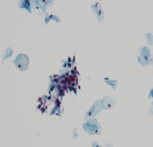
Case 2 from Sue Mehew
Case 3 - Sue Mehew
A 47-year-old first-time screening sample with HPV positivity showed hyperchromatic crowded cell groups (HCGs) and scattered dyskaryotic cells. Reported as severe dyskaryosis, However, histology showed a FIGO 1A1 HPV-associated squamous cell carcinoma.
The case demonstrated that an outcome of invasion is still possible even in the absence of the diagnostic cues such as tumour diathesis, keratinised cells and bizarre cell forms such as fibre/tadpole cells.. Also, the importance of checking around the edge of difficult to visualise HCGs/microbiopsies; focusing up & down through these groups and looking in the background of the preparation for small single cells or small cell clusters that may be easier to interpret.
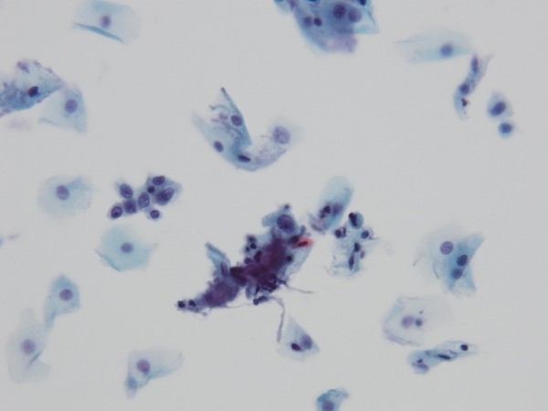
Case 3 from Sue Mehew
Discussion Points
Discussion Points:
- The challenge of diagnosing subtle invasive changes
- The diagnostic value of “fibre”/“tadpole” and keratinised cells
- Importance of context, cytological background, and screening vigilance
- Encouragement of case sharing to build confidence and CPD